How Foot Energy is Dissipated
Presented By. Dr. Bowker
Forces within the foot
There have been a couple of long standing theories as to how energy is dissipated within the hoof. The two conventional schools of thought involve the Depression Theory and the Compression Theory. The Depression Theory is based on force being applied down the limb, through the components of the hoof, and downward to the ground and to a lesser degree outward to the hoof wall.
The Compression Theory is a mirror image of the Depression Theory, with the energy coming up from ground contact and being dissipated up through the limb and to a lesser degree, outward to the hoof wall.
Dr. Bowker decided to utilize engineering testing methodology to determine what forces were at play in the hoof. Through use of transducers, he was able to record the pressure events which occurred within the hoof. What he found was remarkable.
When the maximum force was applied by the hoof of a galloping horse striking hard ground, a negative force was actually generated within the structures above the sole. When Bowker asked an engineer to quantify the negative pressure values obtained in layman's terms, the engineer described the negative pressure as a "Helatious sucking force."
The use of transducers explained what radiographs and fluoroscopic images of feet during the phase of striking the ground and standing on different surfaces were showing. P3 (the pedal bone) was actually moving within the hoof structure.
Navicular issues
Some discussion involved the region where the navicular bone and coffin bone meet along with the intersections of the distal sesamoid impar ligament and deep digital flexor tendon. Research has revealed that radiographic changes which show navicular disease will appear months after the morphology of the navicular bone. Thus this disease is likely a slow process which could allow for the hoof to repair itself somewhat had it been appropriately maintained.
(Sketches will be drawn and scanned in)
Navicular disease seems to be related to stress to fibroblasts in the connective tissues which can be related to improper points of breakover (too long of a toe).
Personal comment added: The issue of stress in fibroblasts would explain why early navicular disease would respond favorably to low intensity light therapy using gallium arsenide lasers or properly modulated infrared LEDs which have been shown to accelerate fibroblast reproduction in the laboratory.
Arterio-venus capillary complexes
Also located at the DSIL / DDFT junction, and of particular interest to me, are arterio-venus capillary complexes; balls of vessels which are quite elaborate but which do not appear to serve any particular tissues. Research shows that they are sensitive to pressures applied to and within the hoof. With the proximity of many nerves, one could conclude that these complexes are sensory mechanisms, allowing the horse to sense the pressure being applied to the ground due to the impact on blood flows. Through their design they also provide a means to absorb some energy when it is redirected and applied to blood flow, thus vascular resistance absorbs a minor portion of the "shock load" to the foot which would otherwise be applied to the hoof components and columnar structures.
Ungual Cartilage
Part of unlocking the mystery relative to the "Helatious sucking force" occurring within the hoof is the understanding of ungual cartilage. Ungual cartilage appeared to be thick in feral horses and in Arabians with similarly durable feet. Instead of being laced with fat, it was very fibrous and resilient. It also completely encased the vasal structures of the arterio-venus capillary complexes.
When the wild horse's foot strikes the ground, pressure is taken up through the bars of the foot, is transmitted by the thick ungual cartilage which rotates the caudal pillars and forces axial projection and blood flow. In short, the structures move in harmony to absorb and dissipate the shock of a galloping horse striking the ground provided that they are in proper alignment and the structures are developed and healthy.
In specimens which had inadequately developed ungual cartilage, the Arterio-venus capillary complexes were exposed and one could surmise were less efficient.
Dr. Bowker also presented a slide from a feral horse which appeared to display a heretofore unnamed ligament which connected the segments of the fully developed ungual cartilage.
(Sketches will be drawn and scanned in)
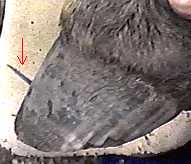
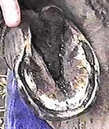
Underrun Heels
Dissection of horses with underrun feet always showed wear under P3. The heels tended to follow the toes. In other words, long toes (too far forward of the apex of the frog) tended to produce underrun (low) heels which affected the internal structures of the hoof and promoted navicular disease.
On feet where there was a pronounced flair on one corner, the opposite diagonal heel would generally be more underrun.
Because of the excessive imbalance of pressure on the hoof capsule, one could predictably expect some signs of laminar damage in these same hooves, so there could be some association between the appearance of some forms of laminitis and eventual navicular disease.
Such hoof deformity also produced observable signs such as interference problems, stumbling and gait faults, probably prior to pathological changes in the navicular area. Thus prevention of navicular disease could be easily improved upon through attainment of a more balanced foot, proper exercise and reasonable observation as to the horse's way of going.
Long Toe / Underrun Heel